Historic Stains (1850 - 1953)
Pre-1850's
Only natural stains were available such as Saffron (which was what Leeuwenhoek used to stain muscle cells)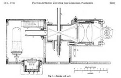
1880's - Paul Ehrlich
Used acidic and basic dyes to identify acidophilic, eosinophilic, basophilic and neutrophilic leukocytes
1904 - August Köhler
Fluorescence UV Microscope
early 1900's - Pappenheim and Unna
Combined methyl green and pyronin to stain nuclei green and cytoplasm red
1925 - Robert Feulgen
Demonstrated that DNA was present in both animal and plant cell nuclei - developed a stoichiometric procedure for staining DNA involving a derivatizing dye, (fuchsin) to a Schiff base
1934 - Andrew Moldavan
Demonstrated use of a suspending fluid in which were blood cells - the measurements were made in a capillary tube using a photoelectric sensor to make extinction measurements
Manuscript: Photo-Electric Technique for the Counting of Microscopical Cells. Andrew Moldavan Montreal, Canada Science 80:188-189, 1934
(1938-1998) -Torbjorn Caspersson - Staining Nucleic acids
1941 - Demonstrated that "nucleic acids, far from being waste products, were necessary prerequisites for the protein synthesis in the cell (published in Naturwissenschaften in January 1941) and that they actively participated in those processes."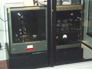
1950 - Demonstrated that both DNA and RNA increase in actively growing cells famous monograph in 1950 "Cell Growth and Cell Function" described nucleic acid and protein metabolism during normal and abnormal growth. These studies were made using a Cadmium spark source for a UV light, and primitive electronic circuits for detection of signals. Used Feulgen stain to stain nuclei.
Manuscript: Torbjorn O. Caspersson, History of the Development of Cytophotometry from 1935 to the present in Analytical and Quantitative Cytology and Histology, pp2-6, 1987.
1941 - Albert H. Coons, Hugh J. Creech and R. Norman Jones
Developed the fluorescence antibody technique - they labeled antipneumococcal antibodies with anthracene allowing them to detect both the organism and the antibody in tissue using UV excited blue fluorescence
Manuscript: Immunological Properties of an Antibody Containing a Fluorescent Group. Albert H. Coons, Hugh J. Creech and R. Norman Jones - Department of Bacteriology and Immunology, Harvard Medical School, and the Chemical Laboratory, Harvard University Proc. Soc. Exp.Biol.Med. 47:200-202, 1941.
1950 - Coons and Melvin H. Kaplan
Conjugated fluorescein with isocyanate - better blue green fluorescent signal - further away from tissue autofluorescence. This method used a very dangerous preparative step using phosgene gas
History of cellular clinical diagnostics
1943 Landmark monograph: Diagnosis of Uterine Cancer by the Vaginal Smear. Authored by George N. Papanicolaou (an anatomist) and Herbert F. Traut (a gynecologist).
Success of the Pap Smear
1941 26,000 deaths per year in the United States due to cancer of the uterus as reported by Papanicolaou and Trout.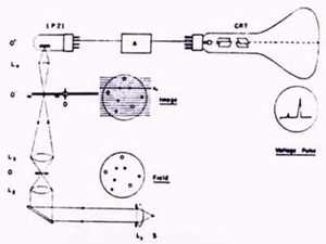
1996 4,900 estimated deaths per year in the United States due to cervical cancer with nearly a 2-fold increase in population in the intervening half century. At least half of these deaths are women who never had a Pap smear.
Pap Testing Improvements
1951 Cytoanalyzer by Airborne instruments of Mineola, New York. Utilized World War II technology. 1980s TICAS and CYBEST; Computer analysis and automated cytology projects. (These pioneering systems proved insufficient for general use).
1944 - Oswald T. Avery
Demonstrated that DNA was the carrier of genetic information.
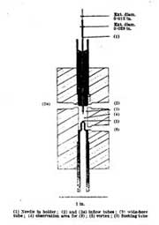
1947 - Gucker
- Developed a flow cytometer for detection of bacteria in aerosols
- Published paper in 1947 (work was done during WWII and was classified).
- Goal was rapid identification of airborne bacteria and spores used in biological warfare
- Instrument: Sheath of filtered air flowing through a dark-field flow illuminated chamber. Light source was a Ford headlamp, PMT detector (very early use of PMT)
1948 - Early Cell Counter
1951 - Early Microfluorometric Scanner Robert Mellors
R.C. Mellors & R. Silver, A microfluorometric scanner for the differential detection of cells: application to exfoliative cytology, Science 104, 1951
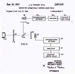
1953 - Two Color Cell Counter Patent
A Device for Counting Small Particles Suspended in a Fluid through a Tube P.J. Crosland-Taylor Bland-Sutton Institute of Pathology Middlesex Hospital, London, W.1. June 17, 1952 Nature 171: 37-38, 1953.
1953 - Watson & Crick
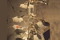
This is a picture of part of the original model built by Watson and Crick at Cambridge in 1953. The work which began with Avery's identification of DNA as the "transforming principle" thus led to research that overturned the old conception of DNA as a repetitive and simple molecule, confirmed DNA's role in genetic transmission, and, with James Watson and Francis Crick's 1953 paper, elucidated its structure.
Blood Cell Counting - Pre-automation
- The hemocytometer was the counting standard until the 1950's.
- The dimension of this device was 3x3x0.1 mm. Typically RBCs were counted using a 1:200 dilution from the 1 x 10⁶/mm3 in whole blood
- Leukocytes (5x10³/mm3) were diluted 1:10 in a lysing reagent and a dye to stain nuclei
- Statistical variation is calculated by the following:
- The standard deviation of a count on n items in n1/2
- Considering no more than 500 cells could be possibly counted manually the standard deviation would therefore be 22
- The coefficient of Variation (CV) is 22/500 or 4.4%
- Add pipetting and dilution errors and it's about 10%
